
AI Agents for Radiology & Post-Deployment Monitoring
Radiology relies on the integration of imaging, clinical data, and timely decision-making, yet most AI tools remain limited to narrow, task-specific predictions. This limits their ability to improve real-world workflows. A key question is how to develop AI systems that understand clinical context, integrate multimodal data, and support radiologic decision-making across the full workflow.
Our lab focuses on building clinical AI agents that can reason over radiologic tasks and take context-aware actions. We study applications such as clinical history summarization, imaging protocol selection, and detection of potential diagnostic errors, with an emphasis on evaluating their impact on efficiency, accuracy, and patient safety in real-world settings.
Our special emphasis is on how to effectively, safely, and efficiently implement AI algorithms and conduct post-deployment continuous monitoring to ensure lasting model performance and safety.
In parallel, we develop the technical foundations needed to support these systems, including simulation environments for realistic training and evaluation, and learning approaches that enable multimodal reasoning over complex clinical workflows. Together, this work aims to establish a scalable framework for end-to-end AI in radiology, enabling more integrated, efficient, and clinically effective workflows.
Quantitative Imaging Research in Cardiothoracic Imaging
Our laboratory has a track record in conducting both clinical research and data science research related to lung cancer and interstitial lung disease imaging. A major emphasis of our research is on developing and validating modern deep learning and quantitative imaging tools to enhance diagnostic accuracy, risk stratification, and prognostication of diseases based on imaging. Our laboratory has numerous in-house algorithms and has also partnered with industry partners to create, validate, and clinically translate advanced imaging and machine learning tools. While CT is our primary modality, we also incorporate other advanced techniques, including molecular imaging and MRI.
In the lung cancer screening space, our team collaborates closely with the lung cancer screening program and tumor boards at UCSF, ZSFG, and the San Francisco VA Medical Center (SFVA), ensuring a comprehensive and multidisciplinary approach to our studies. We have additionally successfully completed a prospective randomized controlled trial (lung cancer trials), such as those focusing on rapid rollover techniques to reduce pneumothorax following CT-guided lung biopsy.
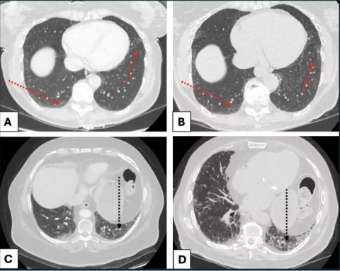
In the interstitial lung disease space, we partner with multidisciplinary physicians in the UCSF Interstitial Lung Disease Clinic to study imaging biomarkers and prognostication in interstitial lung disease. Specifically, our lab has an interest in the early detection and risk stratification of mild fibrosis known as interstitial lung abnormalities (ILA). By leveraging modern hardware and software tools, we seek to maximize diagnostic accuracy, especially at early disease stages, and make a difference in patient management when it can help the most.

Clinical Translation of Mid-Field (0.55T) Lung MRI
In collaboration with UCSF advanced imaging science teams, we are clinically translating cutting-edge hardware and software innovations for cardiothoracic imaging. To this end, we have established ourselves as one of the most active centers in the nation for the pulmonary application of 0.55T MRI, as well as the technical optimizations and innovations surrounding it.
News coverage here.